

One of the strangest measurements in modern cancer metabolism is that many primary solid tumors make ATP more slowly than the healthy tissues they replace. In five mouse tumor models, glycolysis rose as expected, and lower tricarboxylic acid cycle flux dominated the balance, so total ATP production fell. Pancreatic tumors accommodated the deficit by suppressing protein synthesis, while lung metastases recovered higher oxidative flux than their matched primary tumors. A malignant cell can therefore become less energetically productive and more biologically dangerous at the same time.
That anomaly changes the question. The relevant variable is the energetic state a tumor must continuously defend, including production, demand, redox control, substrate exchange, and adaptive reserve. Cancer does change the baseline energetic state of the cell, and “baseline” is a conditional distribution whose value depends on lineage, anatomic location, perfusion, nutrient availability, treatment history, and interactions with immune and stromal cells.
This also changes the therapeutic objective. A useful metabolic therapy should identify the state that sustains a tumor, determine which transitions remain available under pressure, and then collapse the state or close its escape routes. The target is a dynamically maintained configuration, and a pathway label describes only one of its coordinates.
The epistemic setup: beyond the Warburg shorthand
The modern study of cancer metabolism begins with Otto Warburg’s observation that tumors frequently convert glucose to lactate even when oxygen is available. The observation remains foundational. Aerobic glycolysis supplies intermediates for biosynthesis, supports redox management, permits rapid carbon processing, and couples metabolism to growth signaling. It also underlies the clinical utility of fluorodeoxyglucose positron emission tomography.
The Warburg effect became misleading when it was treated as a universal substitution of glycolysis for mitochondrial metabolism. Human tumors often retain active mitochondrial oxidation, use multiple fuels simultaneously, and reorganize nutrient exchange across the tumor microenvironment. Mitochondrial respiration can support proliferation through electron acceptor balance and aspartate synthesis even when ATP itself is sufficiently supplied. Cancer metabolism is therefore a regulatory program that allocates carbon, electrons, and free energy toward survival and reproduction under local constraints.
The consensus has moved toward context dependence. Genetic lesions alter enzyme activity and nutrient preference. Tissue of origin establishes a pre-existing metabolic architecture. Perfusion determines which substrates arrive. Hypoxia and acidity reshape exchange. Therapy changes the fitness landscape and selects resistant states. Immune cells, fibroblasts, and cancer cells partition nutrients according to their own programs, each drawing on the extracellular pool by its own rules.
This richer description creates a measurement problem. Most studies still infer metabolism from transcript abundance, protein abundance, or steady-state metabolite levels. Those measurements reveal components of the machinery. Flux determines what the machinery is doing.
Cancer may occupy energetic attractors
A living cell is an open, driven system maintained far from equilibrium. It continually imports matter and free energy, dissipates heat, exports waste, and uses regulatory feedback to remain inside a viable region of state space. The useful physics is nonlinear dynamics. A stable cellular phenotype behaves like an attractor, a configuration to which the system returns after ordinary perturbations.
We propose that cancer creates lineage- and context-specific energetic attractors. Each attractor is defined by a joint configuration of adenylate balance, glycolytic and oxidative flux, redox ratios, mitochondrial membrane potential, intracellular pH, nutrient exchange, energy expenditure, and spare capacity. The state also includes its accessible transitions, because a tumor capable of switching from glucose oxidation to lactate uptake or fatty-acid oxidation occupies a different therapeutic state from a tumor with the same baseline ATP level and no viable escape route.
A model that measures the full energetic state and its transition capacity should predict response to metabolic perturbation better than the expression of a single enzyme, transporter, or pathway score.
That prediction can be tested in patient-derived systems. Measure the initial state, apply a defined perturbation, observe whether cells die, arrest, differentiate, or switch fuels, and ask which representation best predicted the outcome. Static biomarkers may remain useful, and their value should be judged by their ability to resolve the underlying state.
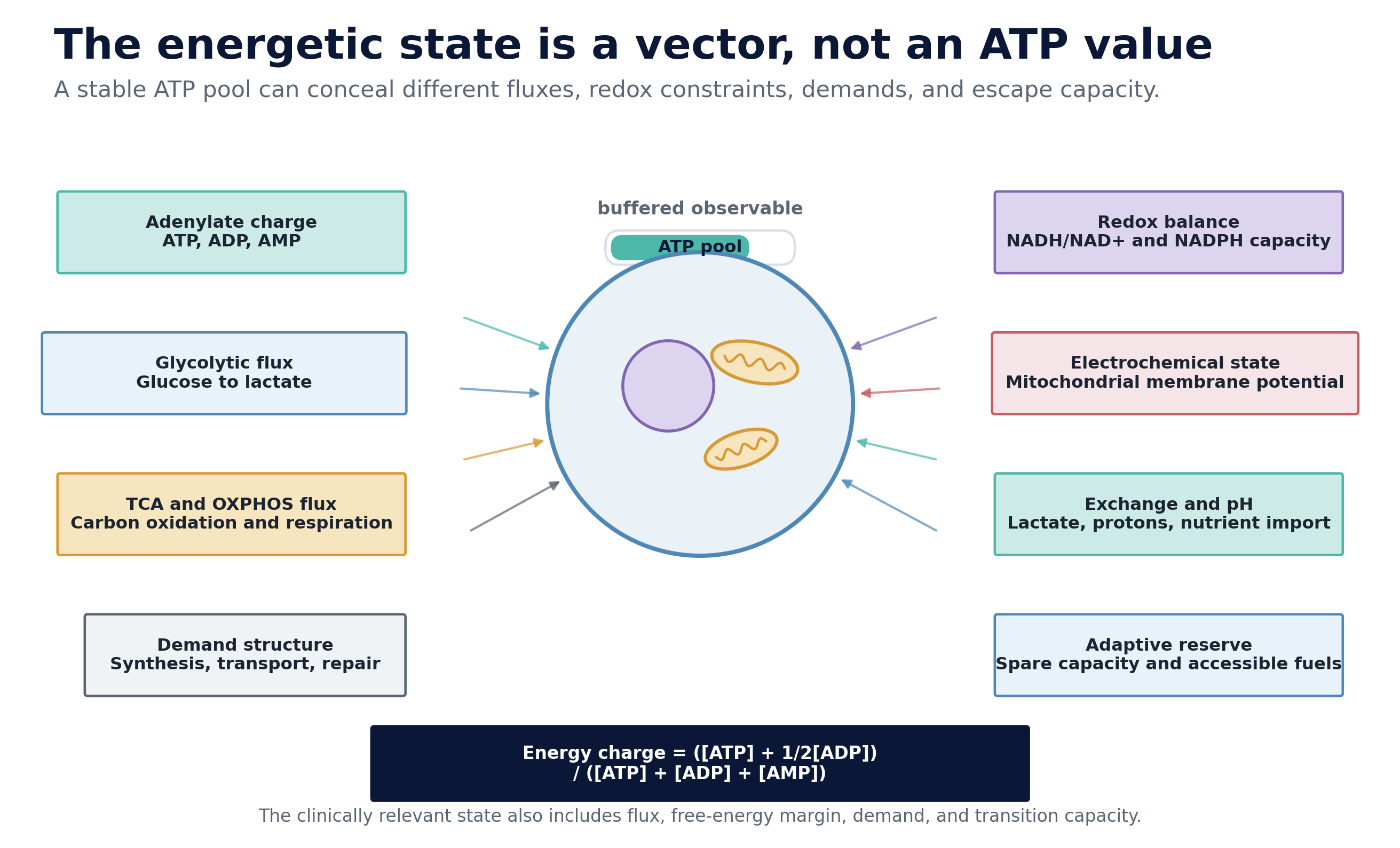
Figure 1. The energetic state is a vector. ATP concentration and adenylate charge are important coordinates, and the same ATP pool can coexist with different fluxes, redox constraints, energy expenditures, membrane potentials, substrate dependencies, and adaptive reserves. The clinically relevant state includes both current coordinates and accessible transitions.
The buffered variable
ATP is indispensable, tightly regulated, and surprisingly poor as a solitary description of cellular energetics. Atkinson’s adenylate energy charge compresses ATP, ADP, and AMP into a normalized measure of the adenylate pool:
Energy charge = ([ATP] + 0.5[ADP]) / ([ATP] + [ADP] + [AMP])
The equation captures a meaningful regulatory variable. It still omits the rate at which ATP is produced and consumed, the free-energy margin of ATP hydrolysis, and the distribution of energetic work across the cell. A stable ATP concentration can be maintained through high production and high consumption, through low production and reduced demand, or through transient depletion buffered by reserve pathways. These states can look similar in a snapshot and respond very differently to treatment.
The 2023 flux study by Bartman and colleagues made this distinction experimentally visible. Primary solid tumors in mice increased glycolysis while lowering TCA-cycle flux enough to reduce total ATP production. Pancreatic tumors compensated by reducing protein synthesis, one of the major energetic costs of differentiated pancreatic tissue. Malignant transformation changed both sides of the ledger. The cells rewired production and shed expensive tissue functions.
This is a general lesson. Cancer can improve fitness by changing what energy is spent on. A malignant cell no longer needs to perform every differentiated function of the tissue it arose from. It can redirect limited energetic capacity toward membrane synthesis, redox buffering, ion gradients, DNA replication, repair, motility, and stress tolerance. The energetic phenotype is therefore partly a demand architecture.
The same reasoning clarifies why mitochondrial respiration cannot be reduced to ATP output. Independent studies in proliferating cells showed that electron transport supports aspartate synthesis by maintaining the electron-acceptor balance required for biosynthesis (Birsoy et al.; Sullivan et al.). A respiratory inhibitor may suppress proliferation through redox and biosynthetic failure before ATP becomes the limiting variable. The therapeutic mechanism and the biomarker should reflect that distinction.
State multiplicity
There is no universal cancer energy state. Intraoperative isotope tracing has shown that human non-small-cell lung cancers often exhibit enhanced glycolysis and glucose oxidation at the same time, with substantial variation within and between tumors. These tumors also oxidize alternative substrates. Subsequent human studies demonstrated that circulating lactate can contribute more carbon to the TCA cycle than glucose in some lung tumors, with MCT1 enabling lactate uptake in experimental models.
Clear-cell renal cell carcinoma occupies a different regime. In patients infused with labeled glucose, these tumors showed enhanced labeling of glycolytic intermediates, suppressed pyruvate dehydrogenase flow, and reduced labeling of the TCA cycle, providing a comparatively strong in vivo example of a glycolytic Warburg phenotype.
The heterogeneity extends beyond tumor type. A 2026 study of fluorodeoxyglucose-negative gastric cancer found evidence that low-glucose-avid tumors can support growth through glutamine-linked gluconeogenic reactions and fatty-acid oxidation. A tumor that appears metabolically quiet through one imaging channel may be active in an unmeasured dimension. Absence of a familiar signal can indicate a different state as readily as an absence of metabolic demand.
Disease stage also matters. In the mouse models studied by Bartman and colleagues, lung metastases had higher TCA-cycle flux than matched orthotopic primary tumors. In melanoma, cells with high MCT1 expression used circulating lactate and managed oxidative stress more effectively; MCT1 inhibition had limited effect on primary subcutaneous tumor growth while reducing circulating tumor cells and metastatic burden in patient-derived xenografts and mouse models. Metastatic competence can therefore be an energetic phenotype with dependencies distinct from those governing primary tumor mass.
These observations support a state-space description. A glycolytic attractor, an oxidative attractor, and a hybrid attractor can all support malignancy. They differ in substrate access, redox logic, microenvironmental exchange, and transition capacity. Histology and genotype constrain the landscape, while environment and treatment determine which basin the cells occupy.
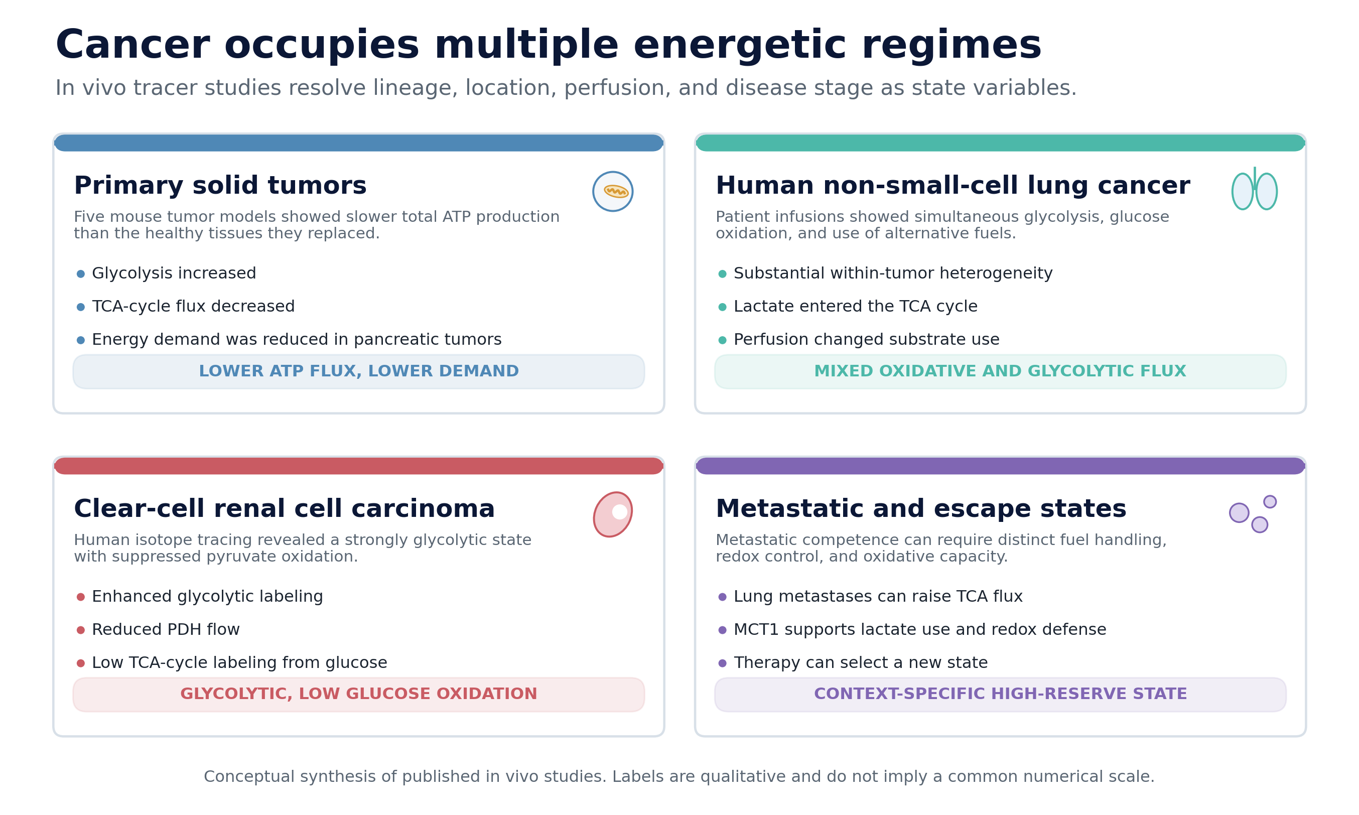
Figure 2. Cancer occupies multiple energetic regimes. The panels summarize qualitative conclusions from in vivo tracer and functional studies. The categories describe distinct regulatory configurations and share no common numerical scale.
Adaptive reserve is part of the disease
A metabolic state is clinically important when it persists under perturbation. The immediate response to pathway inhibition often reveals more than the baseline measurement, because cells expose their reserve fuels, latent transporters, redox buffers, and stress programs only when the main and dominant route is challenged.
This is where many metabolic therapies encounter resistance. Inhibiting glycolysis can increase reliance on mitochondrial oxidation. Inhibiting respiration can increase glycolytic flux, lactate production, and extracellular acidification. Restricting glucose can induce scavenging or the use of alternative carbon sources. These transitions are regulated responses, and they should be modeled as such.
The tumor’s accessible escape routes should therefore be considered part of the target. Two tumors with similar oxygen consumption can have very different vulnerabilities if one can raise glycolysis and the other cannot. Two MCT1-high tumors can respond differently if one also expresses MCT4, which can preserve monocarboxylate transport when MCT1 is inhibited. Two oxidative tumors can differ in their ability to replenish aspartate, regenerate NAD+, or import pyruvate.
The proper experiment resembles a stress test. Baseline measurements define the initial coordinates. A controlled perturbation probes the local curvature of the landscape, revealing how strongly the system returns to its original state, which direction it moves, and where viability fails. In dynamical terms, treatment can shrink an attractor basin, open a transition channel, deepen an alternative basin, or push the system across a viability boundary.
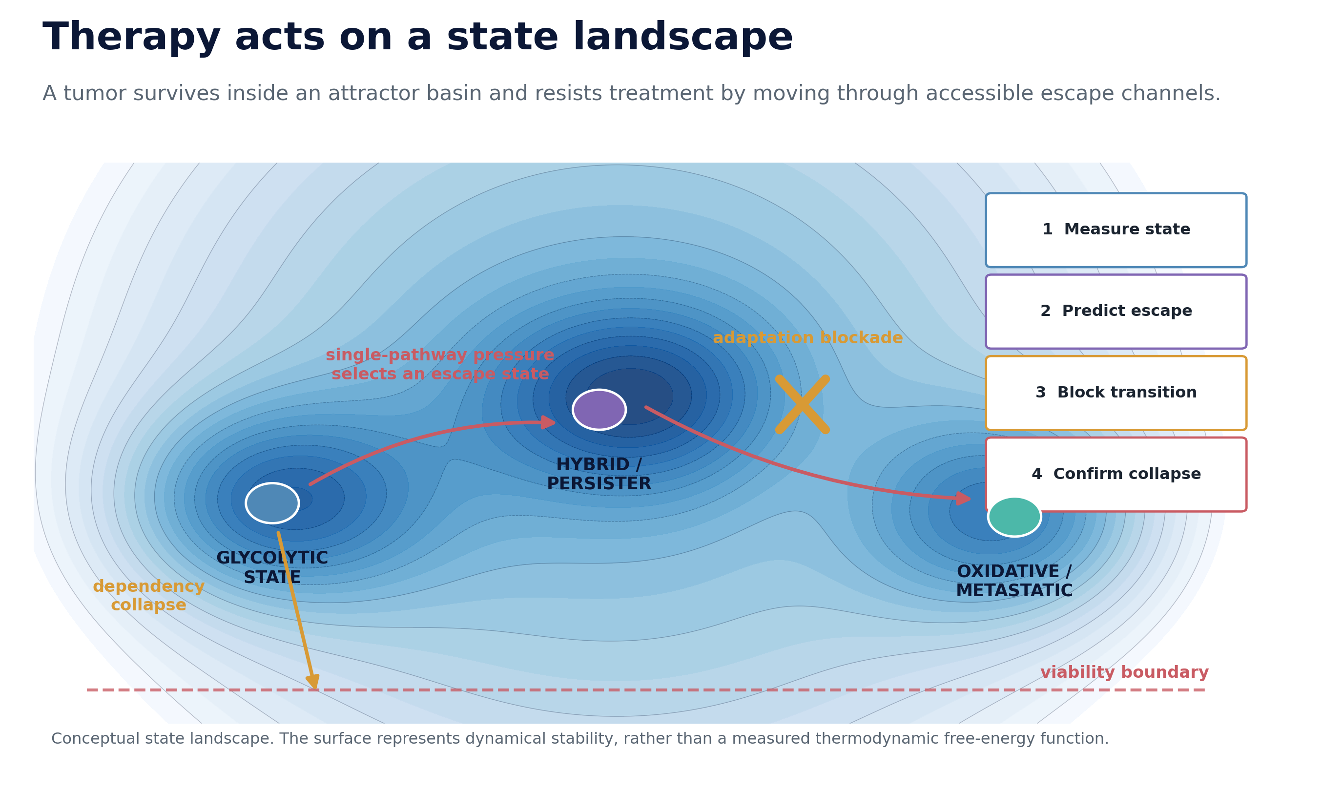
Figure 3. Therapy acts on a state landscape. This is a coarse-grained dynamical schematic of stability; the surface carries no thermodynamic free-energy interpretation. A single-pathway intervention can select an escape state; a successful strategy measures the initial state, predicts the transition, blocks adaptation, and confirms state collapse.
Therapeutic control surfaces
The energetic state can be targeted through several control surfaces. Each requires a different biomarker and fails for a different reason.
1. Collapse a state-specific dependency
A tumor can become unusually dependent on a transporter, enzyme, redox shuttle, or fuel because its other routes are genetically lost, epigenetically suppressed, spatially inaccessible, or already operating near capacity. This is the cleanest form of energetic targeting. The intervention removes a load-bearing component of the state.
The first-in-human study of the MCT1 inhibitor AZD3965 illustrates both the opportunity and the discipline required. The drug achieved target engagement at tolerated doses, and the development strategy focused on tumors with high MCT1 and low MCT4 expression, because MCT4 provides a plausible bypass. On-target toxicities included reversible ocular changes, with additional cardiac and acid-base signals at higher doses. The biology is tractable precisely because dependency and escape can be specified together.
2. Block the adaptive transition
A second strategy targets the escape route itself, leaving the dominant baseline pathway as the first agent’s problem. The combination should follow a mechanistic transition model: inhibit the primary energetic route, identify the compensatory state, and block the regulator or substrate that makes the transition possible. Simultaneous inhibition is not automatically optimal. Sequential timing may exploit a transient state in which the tumor has committed to adaptation and exposed a narrower dependency.
This approach demands functional evidence. A combination justified by two enriched pathway scores remains a correlation. A combination justified by a measured transition, its regulatory driver, and a rescue experiment has a mechanistic architecture.
3. Exploit electrochemical gradients
Mitochondrial membrane potential creates a physical delivery opportunity. Lipophilic cations can accumulate in mitochondria according to the electrochemical gradient, allowing a toxic payload to be concentrated within a selected compartment. Mitochondrially targeted tamoxifen, or MitoTam, used this logic in an early phase I/Ib study across metastatic solid tumors.
The study established clinical feasibility and reported signals of activity in a very small renal-cell carcinoma subgroup. It also produced hematologic toxicity, fever, thromboembolic complications, and serious adverse events, including a fatal stroke. The result is a platform hypothesis awaiting validation. Membrane potential varies across lineages, microenvironments, and treatment states, and normal tissues also depend on mitochondrial electrochemistry. Electrochemical targeting will require direct state measurement and a credible therapeutic window.
4. Target a genetically anchored metabolic lesion
The clearest clinical successes in tumor metabolism have come from states tied to a defined genetic lesion. Mutant IDH enzymes generate the oncometabolite 2-hydroxyglutarate and impose a characteristic epigenetic and differentiation program. In the phase III INDIGO trial, the mutant IDH1/2 inhibitor vorasidenib significantly prolonged progression-free survival in grade 2 IDH-mutant glioma.
Mutant IDH is not a general energy-state target. It is instructive because the metabolic state is anchored by genotype, measurable through a specific metabolite, and linked to a druggable enzyme. The field should seek comparably constrained energetic states whether their origin is regulatory or mutational.
5. Avoid indiscriminate energetic suppression
The failure mode is equally informative. The mitochondrial complex I inhibitor IACS-010759 entered phase I trials with strong preclinical rationale in acute myeloid leukemia and advanced solid tumors. Clinical development encountered a narrow therapeutic index, elevated blood lactate, neurotoxicity, limited target inhibition at tolerated exposure, and little antitumor activity; no recommended phase II dose was established, and both trials were discontinued.
Complex I is a shared requirement across malignant and healthy tissues. A general dependence on respiration does not create selectivity. The relevant question is whether the tumor occupies a state that makes partial inhibition lethal at an exposure healthy tissues can survive. State definition and therapeutic window are the same problem viewed from opposite sides.
Measurement is the binding constraint
The central obstacle in cancer metabolism is measurement quality. Transcriptomics can identify enzymes, transporters, stress responses, and regulatory programs. Proteomics adds abundance and modification state. Metabolomics measures accumulated products and intermediates. Each is informative. None alone determines flux.
Stable-isotope tracing measures the movement of labeled carbon or nitrogen through metabolic networks and has produced some of the most important corrections to textbook models of tumor metabolism. Its clinical use remains logistically demanding, and tissue sampling provides limited spatial and temporal coverage.
Hyperpolarized carbon-13 magnetic resonance imaging offers a complementary route. In a first-in-human prostate cancer study, hyperpolarized pyruvate enabled real-time measurement of pyruvate-to-lactate labeling and identified elevated lactate signal in biopsy-proven tumors. Later work connected lactate labeling to clinically relevant histology, epithelial composition, transporter expression, and spatially resolved transcriptomic features. The method measures a specific exchange process, a single coordinate of the energetic state, and it demonstrates that metabolic dynamics can become a clinical imaging variable.
Spatial metabolomics is beginning to resolve another missing dimension. A 2024 Nature Methods study integrated mass-spectrometry imaging with imaging mass cytometry on the same tissue section, assigning metabolite features to phenotyped cells and revealing metabolic heterogeneity among cancer, stromal, and immune populations. The study also states the core limitation plainly: enzyme and transcript abundance are surrogate markers that may inadequately represent metabolic state.
A robust state measurement architecture will combine several modalities:
Direct flux measurements, including isotope tracing, extracellular flux assays, and dynamic metabolic imaging.
State composition, including metabolites, redox ratios, adenylates, membrane potential, pH, and nutrient exchange.
Regulatory context, including coding and non-coding RNA programs, protein activity, chromatin state, and signaling.
Spatial context, including perfusion, hypoxia, stromal exchange, and immune competition.
Perturbational response, including the direction, speed, and reversibility of state transitions.
Longitudinal observability, allowing the state to be measured repeatedly during treatment.
The final requirement is the least mature. Repeated tumor biopsies are rarely practical. Imaging measures selected reactions and may lack molecular specificity. Plasma offers serial access, and circulating signals mix tumor, immune, vascular, and systemic responses.
A research program for Eigen Bio
For Eigen Bio, energetic-state targeting defines a specific research program for RFM-10B as a transcriptomic state model. RNA should describe the regulatory program that maintains an energetic state, while direct functional assays provide the labels needed to distinguish state control from correlated expression.
The model cannot infer metabolic flux reliably from transcript abundance alone. It should be calibrated against isotope tracing, oxygen-consumption and acidification measurements, metabolite and redox assays, membrane-potential measurements, and spatial data. Patient-derived organoids provide the intervention layer. A predicted dependency can be perturbed under controlled nutrient, oxygen, stromal, and treatment conditions, then evaluated for death, arrest, differentiation, and state switching.
Plasma extracellular-vesicle RNA offers a candidate longitudinal observatory. The working hypothesis is precise: regulatory changes associated with a tumor’s energetic transition are exported into EV-RNA with enough sensitivity and specificity to distinguish tumor response from host response. This remains an experimental question. Matched organoid, tumor, and plasma measurements can test whether the signal is detectable, whether it precedes radiographic change, and whether it identifies the escape route early enough to alter treatment.
The model-lab loop should produce a falsifiable sequence:
Infer the current energetic state and its regulatory program.
Predict the perturbation most likely to destabilize it.
Predict the compensatory transition and the normal-tissue liability.
Test the perturbation in patient-derived organoids with direct functional measurements.
Measure the resulting RNA and EV-RNA state transition.
Compare the predicted and observed trajectory, then update the model.
This is the distinction between a pathway score and a biological world model. A pathway score describes what appears active. A world model predicts what the system will do when acted upon.
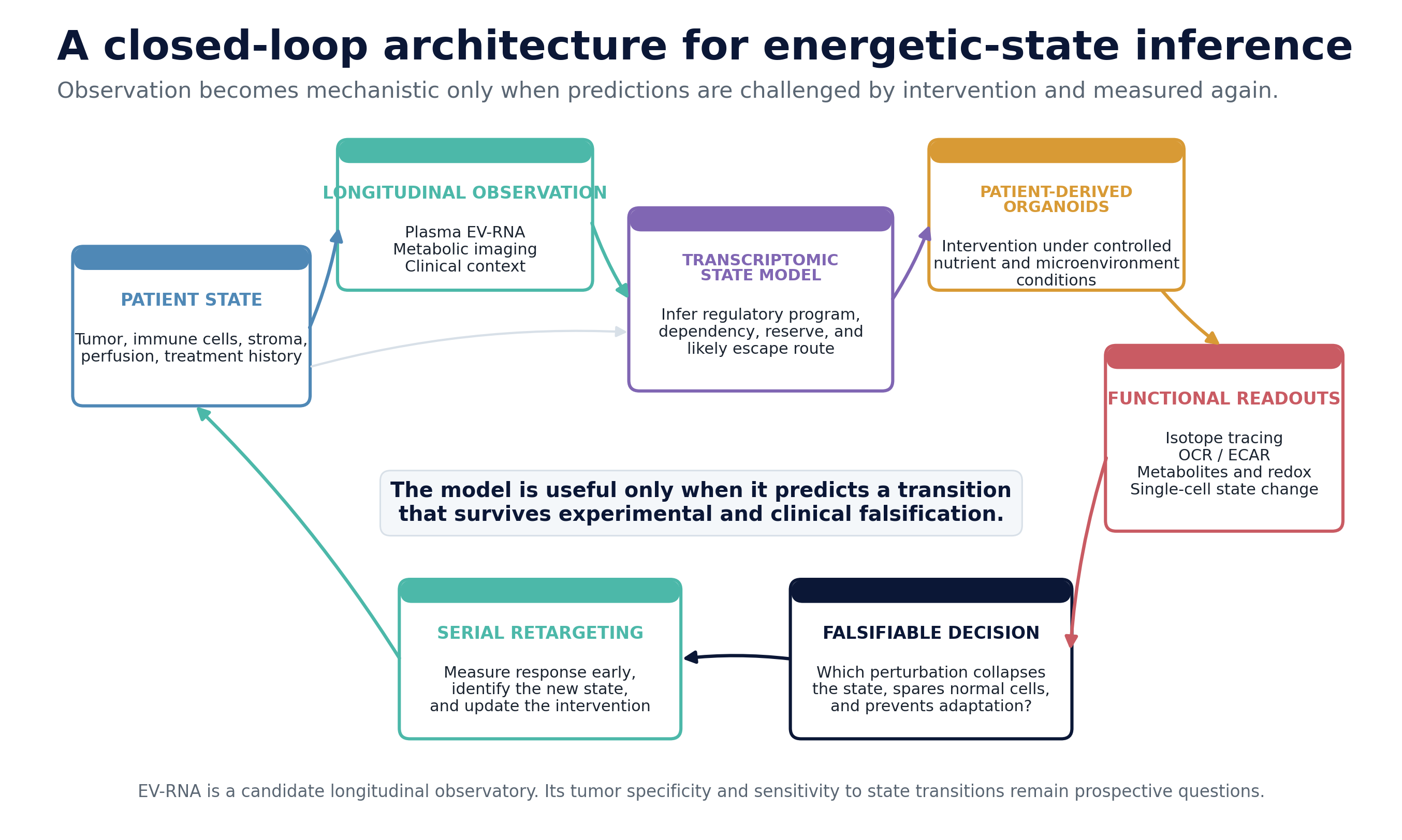
Figure 4. A closed-loop architecture for energetic-state inference. Longitudinal observation, transcriptomic modeling, organoid intervention, and direct functional readouts are connected through prospective falsification. EV-RNA is a candidate observatory whose tumor specificity and transition sensitivity must be established experimentally.
Implications
If the energetic-attractor thesis is correct, metabolic drug development should change in several concrete ways.
Patient selection should be state-based. Expression of the nominal target is insufficient when transport redundancy, substrate availability, or adaptive reserve determines response. Eligibility criteria should encode the dependency, the escape route, and the normal-tissue constraint.
Trials should include a metabolic challenge test. A brief exposure, ex vivo perturbation, or dynamic imaging measurement may reveal vulnerability more effectively than a baseline snapshot. The objective is to observe the direction of movement in state space.
Combinations should be transition-specific. The second agent should block a measured adaptation or exploit the transient state created by the first agent. Combination design becomes a control problem with timing, sequence, and feedback.
On-treatment measurement should occur early. Radiographic response is downstream. A metabolic state can change within minutes to days. Early evidence of state collapse, quiescence, or escape can inform whether continued exposure is biologically rational.
Therapeutic windows should be modeled mechanistically. Tumor selectivity may arise from higher demand, lower reserve, missing redundancy, unusual membrane potential, restricted nutrient access, or a tumor-specific regulatory program. Each source of selectivity can be measured and challenged.
Metastases should be profiled as distinct systems. The energetic state of a metastasis may differ from the primary tumor because the receiving organ changes oxygen, nutrient, and stromal conditions. A treatment chosen from the primary lesion can miss the state that governs lethal disease.
Open questions
Several questions now become experimentally unavoidable.
What is the minimal sufficient state vector? A complete biochemical description is impossible in patients. The practical task is to identify the smallest set of measurements that preserves predictive power across perturbations.
How stable are energetic attractors over time? Some states may persist for months. Others may switch over hours as perfusion, diet, inflammation, or therapy changes. The sampling interval must match the relevant dynamics.
Which state belongs to the cancer cell? Bulk tissue and plasma mix signals from malignant, stromal, immune, endothelial, and systemic compartments. Spatial and single-cell measurements are required to assign causality.
Can EV-RNA resolve a tumor-specific energetic transition? The answer will depend on vesicle origin, RNA cargo selection, tumor burden, clearance, and the magnitude of the regulatory change. Prospective matched experiments can establish the limits.
Where is the tipping point? The most valuable biomarker may be an early sign that the tumor has lost the ability to return to its prior attractor, before cell death or radiographic shrinkage becomes visible.
Can we exploit a state without harming the organism that supports it? Metabolism is shared infrastructure. Precision will come from the tumor’s constrained configuration, its missing redundancies, and its inability to leave a particular state.
Cancer unquestionably alters cellular energetics. The more consequential question is whether we can measure the resulting state precisely enough to identify the transition the tumor cannot survive.
Eigen Bio is a frontier wet AI lab building RFM-10B, a transcriptomic foundation model trained on plasma EV-RNA and patient-derived organoids for mechanistic inference. This post is part of an ongoing series applying tools from physics to the structure of biological discovery.
References
Bartman CR, Weilandt DR, Shen Y, et al. Slow TCA flux and ATP production in primary solid tumours but not metastases. Nature. 2023;614:349-357. doi:10.1038/s41586-022-05661-6
Warburg O. The metabolism of tumors in the body. Journal of General Physiology. 1927;8:519-530. doi:10.1085/jgp.8.6.519
Vander Heiden MG, Cantley LC, Thompson CB. Understanding the Warburg effect: the metabolic requirements of cell proliferation. Science. 2009;324:1029-1033. doi:10.1126/science.1160809
Birsoy K, Wang T, Chen WW, Freinkman E, Abu-Remaileh M, Sabatini DM. An essential role of the mitochondrial electron transport chain in cell proliferation is to enable aspartate synthesis. Cell. 2015;162:540-551. doi:10.1016/j.cell.2015.07.016
Sullivan LB, Gui DY, Hosios AM, Bush LN, Freinkman E, Vander Heiden MG. Supporting aspartate biosynthesis is an essential function of respiration in proliferating cells. Cell. 2015;162:552-563. doi:10.1016/j.cell.2015.07.017
Reinfeld BI, Madden MZ, Wolf MM, et al. Cell-programmed nutrient partitioning in the tumour microenvironment. Nature. 2021;593:282-288. doi:10.1038/s41586-021-03442-1
Atkinson DE. Energy charge of the adenylate pool as a regulatory parameter. Interaction with feedback modifiers. Biochemistry. 1968;7:4030-4034. doi:10.1021/bi00851a033
Hensley CT, Faubert B, Yuan Q, et al. Metabolic heterogeneity in human lung tumors. Cell. 2016;164:681-694. doi:10.1016/j.cell.2015.12.034
Faubert B, Li KY, Cai L, et al. Lactate metabolism in human lung tumors. Cell. 2017;171:358-371.e9. doi:10.1016/j.cell.2017.09.019
Courtney KD, Bezwada D, Mashimo T, et al. Isotope tracing of human clear cell renal cell carcinomas demonstrates suppressed glucose oxidation in vivo. Cell Metabolism. 2018;28:793-800.e2. doi:10.1016/j.cmet.2018.07.020
Liu J, Xia M, Zhao Z, et al. 18F-FDG-PET/CT-negative gastric cancer employs glutamine-based gluconeogenesis and fatty acid oxidation to support tumor growth. Cell Death & Disease. 2026;17:365. doi:10.1038/s41419-026-08662-9
Tasdogan A, Faubert B, Ramesh V, et al. Metabolic heterogeneity confers differences in melanoma metastatic potential. Nature. 2020;577:115-120. doi:10.1038/s41586-019-1847-2
Halford S, Veal GJ, Wedge SR, et al. A phase I dose-escalation study of AZD3965, an oral monocarboxylate transporter 1 inhibitor, in patients with advanced cancer. Clinical Cancer Research. 2023;29:1429-1439. doi:10.1158/1078-0432.CCR-22-2263
Bielcikova Z, Stursa J, Krizova L, et al. Mitochondrially targeted tamoxifen in patients with metastatic solid tumours: an open-label, phase I/Ib single-centre trial. EClinicalMedicine. 2023;57:101873. doi:10.1016/j.eclinm.2023.101873
Mellinghoff IK, van den Bent MJ, Blumenthal DT, et al. Vorasidenib in IDH1- or IDH2-mutant low-grade glioma. New England Journal of Medicine. 2023;389:589-601. doi:10.1056/NEJMoa2304194
Yap TA, Daver N, Mahendra M, et al. Complex I inhibitor of oxidative phosphorylation in advanced solid tumors and acute myeloid leukemia: phase I trials. Nature Medicine. 2023;29:115-126. doi:10.1038/s41591-022-02103-8
Nelson SJ, Kurhanewicz J, Vigneron DB, et al. Metabolic imaging of patients with prostate cancer using hyperpolarized [1-13C]pyruvate. Science Translational Medicine. 2013;5:198ra108. doi:10.1126/scitranslmed.3006070
Sushentsev N, McLean MA, Warren AY, et al. Hyperpolarised 13C-MRI identifies the emergence of a glycolytic cell population within intermediate-risk human prostate cancer. Nature Communications. 2022;13:466. doi:10.1038/s41467-022-28069-2
Nunes JB, Ijsselsteijn ME, Abdelaal T, et al. Integration of mass cytometry and mass spectrometry imaging for spatially resolved single-cell metabolic profiling. Nature Methods. 2024;21:1796-1800. doi:10.1038/s41592-024-02392-6
This is a fascinating, extremely well written and well referenced essay. I greatly enjoyed this Prigoginian/Stuart Kauffman-esque perspective on cancer pathophysiology and therapeutics. I'm a nephrologist, not an oncologist and therefore do not bring any expertise to this discussion, but I have a deep interest in evolutionary biology, per se. Your thesis certainly intersects with the bioenergetic aspects of the atavistic evolutionary model of cancer, which is extremely interesting albeit still subsumed within the fields of theoretical and computational biology. That being said, the models that you presented seem very appealing on theoretical grounds.
In nephrology, the work of Chevalier has been instrumental in bringing evolutionary concepts into the overall discussion of renal pathophysiology, i.e., the transition of living systems from marine to freshwater and then a terrestrial environment has left the human kidney with evolutionary fossils that make us extremely vulnerable to decrements in renal perfusion; scenarios that are seen everyday in hospital ICU's. For example, acute kidney injury due to the various disease processes that cause a decrement in the "effective volume" that perfuses our internal organs, e.g., volume depletion, sepsis, cardiogenic shock, etc. However, as in oncology, primacy, with regards to the tenets of everyday practice is still given to the more traditional "homeostatic" models. Nevertheless, these considerations are a burgeoning source of discussion.
Thankyou for an incredibly informative masterclass in the dynamical systems approach to oncologic bioenergetics and your discussion of the energetic underpinnings of your proposed metabolism based strategy for cancer therapeutics. I am greatly looking forward to your continued discussion of this model which has now greatly informed my personal perspective and working model regarding the role of energetics vis a vis disease pathophysiology, per se. Thank you!